Retina Services
- Services
- Retina Services
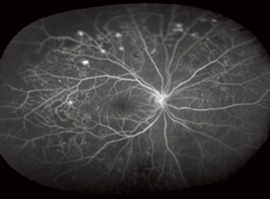
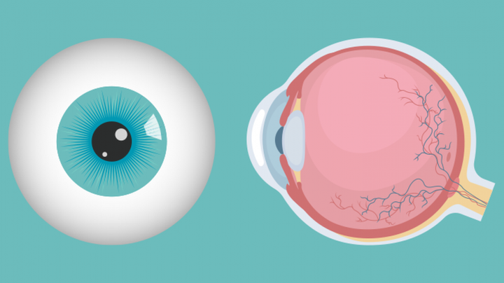
What is Retina?
The retina is a remarkable part of the eye responsible for capturing light and transmitting visual information to the brain. It plays a critical role in our ability to see the world around us. However, several conditions can affect the retina, making it essential to understand these conditions and the treatments available to preserve and restore your vision.
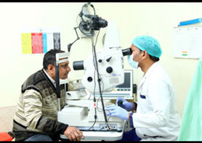
Interested in a Quick 2 Minutes Retina Check Up? - Introducing MIRANTE
The Mirante is not just another diagnostic tool but a significant leap forward in retinal imaging technology. This state-of-the-art machine offers unparalleled imaging capabilities, ensuring precise and detailed retina examinations. Here are the key benefits that Mirante brings to our patients:
Diabetic Screening
Traditional retinal exams required dilation, which could take 1-2 hours. With Mirante, diabetic patients can have their retina status assessed in just 10 seconds. This rapid screening significantly reduces wait time and enhances patient comfort.
Sudden Floaters
Patients experiencing sudden floaters can have an immediate checkup without dilation, providing constant reassurance and timely diagnosis.
LASIK Patients
Patients undergoing LASIK surgery no longer need to wait for lengthy retina checkups before their procedure. Mirante's quick and accurate imaging streamlines the pre-surgery process, making it more efficient and less stressful.
Laser Barrage Follow-Ups
Thanks to Mirante's quick imaging capabilities, follow-up appointments for patients who have undergone laser barrage treatment are now more straightforward and faster.
Myopic Patients
Annual retina checkups for myopic patients, which used to take 2-3 hours, can now be completed in just 10 seconds. This significant reduction in time makes regular checkups more convenient and accessible.
Independence
With Mirante, patients no longer need an attendant or relative to accompany them to their appointments. This independence is particularly beneficial for elderly patients and those with busy schedules.
Post-Dilation Freedom
Traditional dilation can impair near vision and restrict activities such as driving. Mirante eliminates these restrictions, allowing patients to continue their day without interruptions or inconvenience.
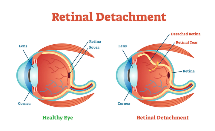
Retinal Detachment: Causes and Treatment
Retinal detachment is a severe eye blinding condition where the retina pulls away from the underlying tissues. This separation can result from various factors, such as:
Torn or Detached Retina
Tears or holes in the retina can lead to detachment. This is often associated with aging or eye injuries.
Vitreous Changes
Changes in the gel-like substance (vitreous) inside the eye can cause retinal detachment.
Myopia
Severe nearsightedness is a risk factor, as it can stretch the retina, making detachment more likely.
Family History
A family history of retinal detachment can increase your risk.
Trauma
A direct injury to the eye can cause retinal detachment.
Treatment for retinal detachment typically involves surgery. Several surgical techniques are available, such as scleral buckle, vitrectomy, and pneumatic retinopexy. The choice of treatment depends on the type and severity of detachment.
Retinal Laser Procedures: Barrage Laser and Green Laser
Retinal laser procedures play a crucial role in the treatment of various retinal conditions. Two common types of retinal lasers are the barrage laser and the green laser:
Barrage Laser
This procedure is used to create a barrier of laser burns around the retinal tear or hole in cases predisposed to retinal detachment. It helps "weld" the retina back in place and prevent further detachment.

Green Laser (Photocoagulation)
Green laser treatment is used to manage conditions like diabetic retinopathy and retinal vein occlusions. It involves directing a focused beam of green laser light onto the retina to seal leaking blood vessels or remove abnormal growths.

Diabetic Retinopathy: Causes and Treatment
Diabetic retinopathy is a diabetes-related eye condition that damages the blood vessels in the retina. It can occur in individuals with both Type 1 and Type 2 diabetes. Causes of diabetic retinopathy include:
High Blood Sugar Levels
High blood sugar levels over time can damage retinal blood vessels.
High Blood Pressure
Elevated blood pressure can contribute to retinal damage.
High Cholesterol Levels
High cholesterol can affect retinal blood vessels.
Smoking
Smoking increases the risk of diabetic retinopathy.
Treatment for diabetic retinopathy may involve laser photocoagulation, anti-VEGF injections (such as bevacizumab, ranibizumab, or aflibercept), or vitrectomy. Early detection and management are crucial in preventing vision loss.

Retinopathy of Prematurity (ROP): Whom to Screen and Treatment
Retinopathy of Prematurity (ROP) is a condition that primarily affects premature infants. It occurs due to abnormal blood vessel development in the retina. High-risk infants, especially those born prematurely or with low birth weight, should be screened for ROP. The severity of ROP will determine the treatment, which may include cryotherapy, laser therapy, or intravitreal injections.
Intravitreal Injections: Indications and Types
Intravitreal injections have revolutionised the treatment of various retinal conditions. These injections deliver medications directly into the vitreous gel of the eye. Common intravitreal injections and their indications include:
Bevacizumab (Avastin)
Used to treat conditions like age-related macular degeneration (AMD), diabetic macular edema, and retinal vein occlusions.
Ranibizumab (Lucentis)
An anti-VEGF drug effective in treating AMD, diabetic retinopathy, and macular edema.
Aflibercept (Eylea)
Another anti-VEGF medication used for AMD and diabetic retinopathy.
Brolucizumab (Beovu)
Newer anti-VEGF molecule for the treatment of AMD and diabetic macular edema.
Ozurdex (Dexamethasone implant)
These are used to treat macular edema due to various causes.
These injections have been instrumental in stabilizing and even improving vision in patients with retinal conditions, offering hope for many who may have otherwise faced significant vision loss.
In conclusion, retinal health is of paramount importance for maintaining good vision. If you have concerns about your retina or experience any vision changes, consult with an eye specialist who can provide a thorough examination and recommend appropriate treatments, including laser procedures and intravitreal injections when necessary. Early diagnosis and intervention are key to preserving your sight and enjoying a life of visual clarity and well-being.
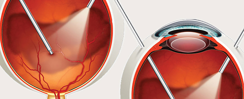
Vitrectomy Surgery: Shedding Light on Advanced Eye Care
Vitrectomy surgery is a remarkable procedure that plays a crucial role in preserving and restoring vision in individuals with complex eye conditions. This advanced surgical technique is particularly effective in treating a range of retinal and vitreous issues. At AcuraVision Clinics, we take pride in offering state-of-the-art eye care solutions, including the cutting-edge technology of The Constellation, by Alcon, which elevates our commitment to exceptional patient care.
Indications for Vitrectomy Surgery
Vitrectomy surgery is recommended for a variety of eye conditions, especially when more conservative treatments have proven insufficient. Some of the primary indications for vitrectomy include:
Retinal Detachment
In cases of retinal detachment, vitrectomy can help reattach the retina by removing the vitreous gel that might be pulling it away from the eye's inner wall. This is vital in preventing vision loss.
Diabetic Retinopathy
For individuals with advanced diabetic retinopathy, blood vessel abnormalities in the retina can lead to severe vision problems. Vitrectomy is often performed to clear the blood from the vitreous gel.
Macular Holes and Epiretinal Membranes
Vitrectomy is used to treat macular holes and epiretinal membranes by removing the gel to allow the retina to lie flat.
Vitreous Hemorrhage
In cases of vitreous hemorrhage due to various causes, including trauma or retinal vessel bleeding, vitrectomy can clear the blood from the eye to restore vision.
Infections and Inflammation
Severe eye infections or inflammation within the vitreous may necessitate a vitrectomy to remove infected tissue and promote healing.
The Constellation by Alcon
AcuraVision Clinics is proud to offer our patients access to the latest advancements in eye care. The Constellation, by Alcon, is a state-of-the-art vitrectomy system that sets new standards in precision, safety, and effectiveness. This cutting-edge technology allows our skilled surgeons to perform vitrectomy procedures with unparalleled precision and minimal disruption to the eye.
With its advanced micro-incision capabilities and advanced visualization, The Constellation system empowers our surgeons to navigate the delicate structures within the eye with great accuracy. This results in reduced surgery time, faster recovery, and improved outcomes for our patients.
In summary, vitrectomy surgery is a remarkable advancement in the field of eye care, providing hope and healing for individuals with a range of retinal and vitreous conditions. Whether you are dealing with retinal issues, diabetic retinopathy, or other vitreous conditions, you can trust AcuraVision Clinics for outstanding care and exceptional results.
Why Choose AcuraVision for Retina Care?
- State-of-the-art Mirante technology for quick 10-second retina checkups without dilation
- The Constellation by Alcon - advanced vitrectomy system for precision surgery
- Board-certified retina specialists with extensive experience
- Comprehensive treatment options including laser procedures and intravitreal injections
- Advanced diagnostic and surgical equipment
- Comprehensive care for complex retinal conditions
- Emergency services for retinal detachments and urgent conditions
- Early detection and intervention to preserve and restore vision